
A Lightweight Explainable AI Framework for Early Brain Tumor Detection Using EEG Functional Connectivity
DOI:
https://doi.org/10.54327/set2026/v6.i2.368Keywords:
brain tumor detection; electroencephalography; graph neural networks; functional connectivity; explainable artificial intelligence; lightweight deep learningAbstract
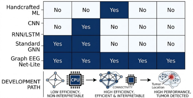
Early identification of brain tumors is an important technique for timely clinical assessment, but magnetic resonance imaging (MRI) and computed tomography (CT) remain the primary modalities for anatomical confirmation and tumor localization. Electroencephalography (EEG) provides a non-invasive and comparatively accessible complementary modality that can capture functional alterations in neural activity. This study presents Graph EEG Net-Lite, a lightweight and explainable graph neural network for auxiliary brain tumor screening using resting-state EEG functional connectivity. EEG recordings from 50 subjects, comprising 25 participants with radiologically confirmed brain tumors and 25 neurologically healthy controls, were represented as functional connectivity graphs using 19 EEG channels. Signals were filtered using a zero-phase fourth-order Butterworth band-pass filter from 0.5 to 45 Hz and a 50 Hz notch filter, segmented into 4-s non-overlapping epochs, and represented using relative spectral power and phase-locking value (PLV)-based connectivity. Graph EEG Net-Lite employs two Chebyshev graph convolution blocks with 64- and 32-dimensional hidden representations, followed by global mean pooling and a shallow classifier. Under subject-independent evaluation, the model achieved 96.2% accuracy, 96.8% sensitivity, 95.5% specificity, 95.9% F1-score, and an AUC of 0.987. The model used 74.8% fewer parameters and required 61.3% less training time than the standard GNN baseline. GNNExplainer identified informative contributions from frontal, temporal, parietal, and occipital electrode regions. The findings support the feasibility of lightweight graph-based EEG analysis as an auxiliary screening and decision-support approach. At the same time, MRI and CT remain necessary for clinical confirmation and anatomical characterization.
Downloads
Downloads
Published
Data Availability Statement
The author(s) have made their research data available for access by readers.Issue
Section
Categories
License
Copyright (c) 2026 Haider TH. Salim AlRikabi, Mohand Lokman Al Dabag, Razan Abdulhammed, Hussain Ali Mutar, Ibtihal Razaq Niama ALRubeei, Shaima Miqdad Mohamed Najeeb

This work is licensed under a Creative Commons Attribution 4.0 International License.
Authors are permitted and encouraged to post their work online (e.g., in institutional repositories or on their website, social networking sites, etc).


 © SET Journal